Your Medical Advices: Medical Articles
Showing posts with label Medical Articles. Show all posts

Monday, October 6, 2014

Circuitry of Cardiovascular System

The steps in one complete circuit through the cardiovascular system are shown in figure. The cycled numbers in the figure correspond with the steps described here: 
A schematic diagram showing the circuitry of the cardiovascular system
  1. Oxygenated blood fills the left ventricle.  Blood that has been oxygenated in the lungs returns to the left atrium via the pulmonary vein. This blood then flows from the left atrium to the left ventricle through the mitral valve (the AV valve of the left heart).
  2. Blood is ejected from the left ventricle into the aorta.  Blood leaves the left ventricle through the aortic valve (the semilunar valve of the left side of the heart), which is located between the left ventricle and the aorta. When the left ventricle contracts, the pressure in the ventricle increases, causing the aortic valve to open and blood to be ejected forcefully into the aorta. (As noted previously, the amount of blood ejected from the left ventricle per unit time is called the cardiac output.) Blood then flows through the arterial system, driven by the pressure created by contraction of the left ventricle.
  3. Cardiac output is distributed among various organs.  The total cardiac output of the left heart is distributed among the organ systems via sets of parallel arteries. Thus, simultaneously, 15% of the cardiac output is delivered to the brain via the cerebral arteries, 5% is delivered to the heart via the coronary arteries, 25% is delivered to the kidneys via the renal arteries, and so forth. Given this parallel arrangement of the organ systems, it follows that the total systemic blood flow must equal the cardiac output.
  4. Blood flow from the organs is collected in the veins.  The blood leaving the organs is venous blood and contains waste products from metabolism, such as carbon dioxide (CO2). This mixed venous blood is collected in veins of increasing size and finally in the largest vein, the vena cava. The vena cava carries blood to the right heart.
  5. Venous return to the right atrium.  Because the pressure in the vena cava is higher than in the right atrium, the right atrium fills with blood, the venous return. In the steady state, venous return to the right atrium equals cardiac output from the left ventricle. 
  6. Mixed venous blood fills the right ventricle.  Mixed venous blood flows from the right atrium to the right ventricle through the AV valve in the right heart, the tricuspid valve.
  7. Blood is ejected from the right ventricle into the pulmonary artery.  When the right ventricle contracts, blood is ejected through the pulmonic valve (the semilunar valve of the right side of the heart) into the pulmonary artery, which carries blood to the lungs. Note that the cardiac output ejected from the right ventricle is identical to the cardiac output that was ejected from the left ventricle. In the capillary beds of the lungs, oxygen (O2) is added to the blood from alveolar gas, and CO2 is removed from the blood and added to the alveolar gas. Thus, the blood leaving the lungs has more O2 and less CO2 than the blood that entered the lungs.
  8. Blood flow from the lungs is returned to the heart via the pulmonary vein.  Oxygenated blood is returned to the left atrium via the pulmonary vein to begin a new cycle.
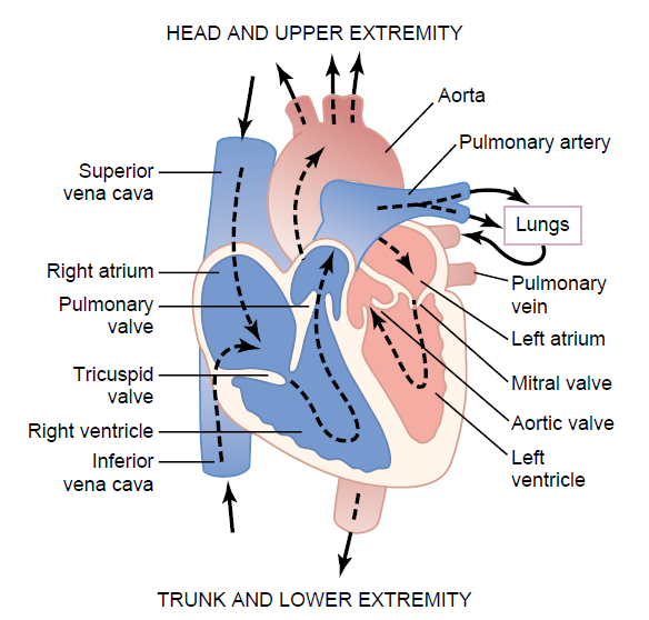
Structure of the heart, and course of blood flow through the heart
chambers and heart valves.


    Gynecologic History

    General
    • Name, age and occupation
    • A brief statement of the general nature and duration of the main complaints (try to use the patient’s own words rather than medical terms at this stage)

    History of presenting complaint
    • This section should focus on the presenting complaint, e.g. menstrual problems, pain, subfertility, urinary incontinence, etc. The detailed questions relating to each complaint are covered in more detail in the relevant chapters, but there are certain important aspects of a gynaecological history that should always be enquired about.
    Menstrual history
    • Age of menarche
    • Usual duration of each period and length of cycle (usually written as mean number of days of bleeding over usual length of full cycle, e.g. 5/28)
    • First day of the last period
    • Pattern of bleeding: regular or irregular and length of cycle
    • Amount of blood loss: more or less than usual, number of sanitary towels or tampons used, passage of clots or flooding
    • Any intermenstrual or post-coital bleeding
    • Any pain relating to the period, its severity and timing of onset
    • Any medication taken during the period (including over-the-counter preparations).
    Previous gynaecological history
    • This section should include any previous gynaecological treatments or surgery.
    Previous obstetric history
    • Number of children with ages and birth weights.
    • Any abnormalities with pregnancy, labour or the puerperium
    • Number of miscarriages and gestation at which they occurred
    • Any terminations of pregnancy with record of gestational age and any complications.
    Previous medical history
    • Any serious illnesses or operations with dates
    • Family history.
    Enquiry about other systems
    • Appetite, weight loss, weight gain
    • Bowel function (if urogynaecological complaint, more detail may be required)
    • Bladder function (if urogynaecological complaint, more detail may be required).
    • Enquiry of other systems
    Social history
    • Sensitive enquiry should be made about the woman’s social situation including details of her occupation, who she lives with, her housing and whether or not she’s in a stable relationship.
    • A history regarding smoking and alcohol intake should also be obtained. Any pertinent family or other relevant social problems should be briefly discussed. If admission and surgery are being contemplated it’s necessary to establish what support she has at home, particularly if she is elderly or frail.

    Heart

    Cardiac orientation

    The general shape and orientation of the heart are that of a pyramid that has fallen over and is resting on one of its sides . Placed in the thoracic cavity, the apex of this pyramid projects forward, downward, and to the left, whereas the base is opposite the apex and faces in a posterior direction (Fig. 1) . The sides of the pyramid consist of:
    • diaphragmatic (inferior) surface on which the pyramid rests,
    • an anterior (sternocostal) surface oriented anteriorly,
    • a right pulmonary surface, and
    • a left pulmonary surface.
    Fig. 1 Base of the Heart

    Because the great veins enter the base of the heart, with the pulmonary veins entering the right and left sides of the left atrium and the superior and inferior venae cavae at the upper and lower ends of the right atrium, the base of the heart is fixed posteriorly to the pericardia! wall, opposite the bodies of vertebrae TV to TVIII (TVI to TIX when standing) . The esophagus lies immediately posterior to the base.

    From the base the heart proj ects forward, downward, and to the left, ending in the apex. The apex of the heart is formed by the inferolateral part of the left ventricle (Fig. 2) and is positioned deep to the left fifth intercostal space, 8-9 em from the midsternal line.
    Fig. 2 Anterior Surface of the Heart

    Surfaces of the heart

    The anterior surface faces anteriorly and consists mostly of the right ventricle, with some of the right
    atrium on the right and some of the left ventricle on the left (Fig. 2 ).

    The heart in the anatomical position rests o n the diaphragmatic surface, which consists of the left ventricle
    and a small portion of the right ventricle separated by the posterior interventricular groove (Fig. 3) . This surface faces inferiorly, rests on the diaphragm, is separated from the base of the heart by the coronary sinus, and extends from the base to the apex of the heart.

    The left pulmonary surface faces the left lung, is broad and convex, and consists of the left ventricle and a
    portion of the left atrium (Fig. 3).

    The right pulmonary surface faces the right lung, is broad and convex, and consists of the right atrium
    (Fig. 3).
    Figs. 3 Diaphragmatic surface of the Heart 

    Margins and Borders

    Some general descriptions of cardiac orientation refer to right, left, inferior (acute), and obtuse margins:
    • The right and left margins are the same as the right and left pulmonary surfaces of the heart.
    • The inferior margin is defined as the sharp edge between the anterior and diaphragmatic surfaces of the heart (Figs. 2)-it is formed mostly by the right ventricle and a small portion of the left ventricle near the apex.
    • The obtuse margin separates the anterior and left pulmonary surfaces (Fig. 2)-it is round and extends from the left auricle to the cardiac apex (Fig. 2) , and is formed mostly by the left ventricle and superiorly by a small portion of the left auricle.
    External sulci

    Internal partitions divide the heart into four chambers (i.e. , two atria and two ventricles) and produce surface or external grooves referred to as sulci.
    • The coronary sulcus circles the heart, separating the atria from the ventricles {Fig. 4) . As it circles the heart, it contains the right coronary artery, the small cardiac vein, the coronary sinus, and the circumflex branch of the left coronary artery.
    • The anterior and posterior interventricular sulci separate the two ventricles-the anterior interventricular sulcus is on the anterior surface of the heart and contains the anterior interventricular artery and the great cardiac vein, and the posterior interventricular sulcus is on the diaphragmatic surface of the heart and contains the posterior interventricular artery and the middle cardiac vein.


    CARDIAC CHAMBERS

    The heart functionally consists of two pumps separated by a partition (Fig.1). The right pump receives deoxygenated blood from the body and sends it to the lungs. The left pump receives oxygenated blood from the lungs and sends it to the body. Each pump consists of an atrium and a ventricle separated by a valve.

    The thin-walled atria receive blood coming into the heart, whereas the relatively thick-walled ventricles pump
    blood out of the heart.
    More force is required to pump blood through the body than through the lungs, so the muscular wall of the left ventricle is thicker than the right.
    Fig. 1 The Heart has two pumps


    Right Atrium

    In the anatomical position, the right border o f the heart is formed by the right atrium. This chamber also contributes to the right portion of the heart's anterior surface.
    Blood returning to the right atrium enters through one of three vessels. These are:
    • the superior and inferior venae cavae, which together deliver blood to the heart from the body; and
    • the coronary sinus, which returns blood from the walls of the heart itself.
    The superior vena cava enters the upper posterior portion of the right atrium, and the inferior vena cava and
    coronary sinus enter the lower posterior portion of the right atrium.
    From the right atrium, blood passes into the right ventricle through the right atrioventricular orifice. This opening faces forward and medially and is closed during ventricular contraction by the tricuspid valve.

    The interior of the right atrium is divided into two continuous spaces. Externally, this separation is indicated by a shallow, vertical groove (the sulcus terminalis cordis) , which extends from the right side of the opening of the superior vena cava to the right side of the opening of the inferior vena cava. Internally, this division is indicated by the crista terminalis (Fig. 2) , which is a smooth, muscular ridge that begins on the roof of the atrium just in front of the opening of the superior vena cava and extends down the lateral wall to the anterior lip of the inferior vena cava.
    The space posterior to the crista is the sinus of venae cavae and is derived embryologically from the right horn of the sinus venosus. This component of the right atrium has smooth, thin walls, and both venae cavae empty into this space.

    The space anterior to the crista, including the right auricle, is sometimes referred to as the atrium proper.
    This terminology is based on its origin from the embryonic primitive atrium. Its walls are covered by ridges called the musculi pectinati (pectinate muscles), which fan out from the crista like the "teeth of a comb . " These ridges are also found in the right auricle, which is an ear-like, conical, muscular pouch that externally overlaps the ascending aorta.

    An additional structure in the right atrium is the opening of the coronary sinus, which receives blood
    from most of the cardiac veins and opens medially to the opening of the inferior vena cava. Associated with these openings are small folds of tissue derived from the valve of the embryonic sinus venosus (the valve of the coronary sinus and the valve of inferior vena cava, respectively) . During development, the valve of the inferior vena cava helps direct incoming oxygenated blood through the foramen ovale and into the left atrium.

    Separating the right atrium from the left atrium is the interatrial septum, which faces forward and to the right
    because the left atrium lies posteriorly and to the left of the right atrium. A depression is clearly visible in the septum just above the orifice of the inferior vena cava. This is the fossa ovalis (oval fossa) , with its prominent margin, the limbus fossa ovalis (border of the oval fossa).

    The fossa ovalis marks the location of the embryonic foramen ovale, which is an important part of fetal circulation. The foramen ovale allows oxygenated blood entering the right atrium through the inferior vena cava to pass directly to the left atrium and so bypass the lungs, which are nonfunctional before birth.

    Finally, numerous small openings-the openings of the smallest cardiac veins (the foramina of the venae
    cordis minimae)-are scattered along the walls of the right atrium. These are small veins that drain the myocardium directly into the right atrium.

    Tuesday, September 9, 2014

    Gram-Positive Bacteria


    General Characteristics

          -Gram-positive bacteria have a highly cross-linked, multilayered (and usually thick) peptidoglycan    cell wall which traps the large Gram crystal violet-iodine complex staining them deep purple.

          -Teichoic acids are linked to either the cytoplasmic membrane (the lipoteichoic acids) or to the cell wall peptidoglycan. Teichoic acids are unique to Gram-positive bacteria and play roles in adherence and triggering Gram-positive shock as the cell wall is broken down.
          -A variety of cell surface proteins are present, which are organism specific.
          -Gram-positive bacteria have no outer membrane and therefore no hydrophobic barrier to limit access of larger antibiotics to the peptidoglycan.

              Major genera of Gram-positive bacteria


              • Genus: Staphylococcus. Common name: Staphylococci
                • Features
                  • Gram-positive cocci generally in tight grapelike clusters or, in specimens, as singlets, pairs, or short chains as well as clusters.
                  • Catalase-positive, breaking down hydrogen peroxide into water and oxygen.
                  • Facultative anaerobes producing energy more efficiently aerobically.
                  • Haloduric (salt-tolerant).
                  • Speciated medically on the basis of coagulase and hemolysis.
              • Genus: Streptococcus. Common name: Streptococci
                • Features
                  • Gram-positive cocci in chains or pairs. They tend to be oval.
                  • Distinguished from staphylococci on the basis of the catalase test. Streptococci are catalase-negative, aerotolerant anaerobes that grow in full oxygen but ferment both in the presence and absence of oxygen.
                  • Subdivided or speciated by three different systems: serology, hemolysin production, and biochemical properties.
                    • Serology using Lancefield’s antibodies to cell wall carbohydrates
                      • Streptococci that are positive for these carbohydrates are classified into Lancefield’s serogroups (e.g., Group A strep). There are now more than 20 groups.
                      • Bacteria that have these cell wall carbohydrates produce a pyogenic reaction.
                      • Some alpha-hemolytic streptococci including Strep. pneumoniae and the viridans streptococci lack these cell wall carbohydrates. They are not grouped using Lancefield’s antibodies and are not pyogenic.
                    • Hemolysin testing
                    • Biochemical tests
                  • Streptococci are mainly opportunists but can cause disease in debilitated patients or if they gain entry into the body.
              • Genus: Enterococcus
                • Features
                  • Catalase-negative, facultative anaerobes fermenting even in full oxygen; Streptococcal family.
                  • Alpha-hemolytic or nonhemolytic, Gram-positive cocci in chains that have the Group D streptococcal cell wall carbohydrate.
                  • Part of the normal human gastrointestinal (GI) flora.
                  • Tolerant of high concentrations of bile salts and NaCl.
                  • Have a high level of drug resistance that continues to increase due to efficient acquisition of plasmid transposon genes for drug resistance.
              • Genus: Peptostreptococcus
                • Features
                  • Peptostreptococci are obligate anaerobic streptococci
                  • They are part of the normal flora of the oral, intestinal, and genitourinary tracts
              • Genus: Bacillus
                • Features
                  • Gram-positive, spore-forming rods that may form chains. Although spores survive for decades in dry environments, they quickly germinate in rich moist conditions (e.g., a macrophage) and transform into metabolically active vegetative cells.
                  • They are aerobes (or facultative anaerobes) and grow well in ambient air.
                  • They cause anthrax (Bacillus anthracis) and food poisoning (Bacillus cereus).
              • Genus: Clostridium. Common name: Clostridia
                • Features
                  • Anaerobic, Gram-positive, spore-forming rods that can form chains.
                  • Cause botulism (Cl. botulinum) characterized by a flaccid paralysis; gas gangrene and food poisoning (Cl. perfringens); and tetanus (Cl. tetani), characterized by rigid spasms.
              • Genus: Listeria. Listeria monocytogenes is the only human pathogen
                • Features
                  • Short Gram-positive, non-spore-forming rod showing weak beta-hemolysis on blood agar.
                  • Motile in broth by tumbling. (They lack forward movement.)
                  • Facultative intracellular pathogens; they move from cell to cell by actin polymerization, which may propel the bacterium directly into an adjoining cell without exposure to extracellular milieu.
                  • Grows in the cold and, unlike most non-spore-forming pathogens, survives in the environment.
                  • Listeria monocytogenes is found in animal feces, rotting vegetation, and occasionally in soft cheeses, deli meats, and cabbage.
                  • Causes mild gastroenteritis, as well as septicemia in pregnant women, leading to potential fetal septicemia or meningitis, and may cause meningitis in immunocompromised patients.
              • Genus: Erysipelothrix
                • Features
                  • Aerobic Gram-positive, non-spore-forming rods.
                  • Found in animals and rotting organic material; entry is through traumatic implantation.
                  • Cause cutaneous erysipeloid primarily in fishmongers, butchers, and veterinarians.
              • Genus: Corynebacterium
                • Features
                  • Club-shaped, Gram-positive non-spore-forming bacteria.
                  • Found in Chinese character-like arrangement of cells.
                  • Aerobic and nonmotile.
                  • Part of normal flora; the non-toxin-producing corynebacteria found in the normal microbiota are called diphtheroids.
                  • Tox+ C. diphtheriae causes diphtheria, and Corynebacterium jeikeium causes infections via catheters and foreign bodies in immunocompromised hosts.
              • Genus: Actinomyces
                • Features
                  • Anaerobic, Gram-positive rods with some branching; nonmotile.
                  • Found in crevices between teeth and gums and female genital tract.
                  • Not acid-fast.
                    • These bacteria are related to mycobacteria.
                    • They have a similar cell wall but lack the extremely long chain fatty acids found in the mycobacterial cell wall. They have shorter fatty acid chains.
                  • Cause cervicofacial or pelvic infections following trauma that has resulted in necrotic tissue; colonies formed in tissue are sometimes described as “sulfur” granules.
              • Genus: Nocardia.
                • Features
                  • Gram-positive filamentous bacteria breaking up into rods.
                  • Aerobic soil organism.
                  • Often described as weakly or partially acid-fast as the slide will show some areas where the cells retain some of the carbol-fuchsin (hot pink red color).
                  • Related to mycobacteria—have a cell wall with shorter chain mycolic acids; are somewhat resistant to drying, so they are transmitted in dust.
                  • Cause tuberculosis-like (but not contagious) bronchopulmonary disease in immunocompromised patients.

              Monday, September 8, 2014

              Inflammation

              The innate immune system is programmed to respond to damage to the body, whether the damaged tissue is septic or sterile. That response rapidly initiates an interactive system of humoral (soluble in the blood) and cellular systems, called inflammation. Inflammation is the first response to injury.
              The inflammatory response:

              • occurs in tissues with a blood supply (vascularized)
              • is activated rapidly (within seconds) after damage occurs
              • depends on the activity of both cellular and chemical components
              • is nonspecific


              The classic symptoms of acute inflammation include redness (erythema), heat, swelling, pain, and loss of function. Microscopic inflammatory changes occur within seconds in the microcirculation (arterioles, capillaries, and venules) near the site of an injury and include the following processes:

              • Vasodilation (increased size of the blood vessels), which causes slower blood velocity and increases blood flow to the injured site
              • Increased vascular permeability (the blood vessels become porous from contraction of endothelial cells) and leakage of fluid out of the vessel (exudation), causing swelling (edema) at the site of injury; as plasma moves outward, blood in the microcirculation becomes more viscous and flows more slowly, and the increased blood flow and increasing concentration of red cells at the site of inflammation cause locally increased redness (erythema) and warmth.
              • White blood cell adherence to the inner walls of vessels and their migration through enlarged junctions between the endothelial cells lining the vessels into the surrounding tissue.
              There are several benefits of inflammation, including the following:
              • Prevents infection and further damage by contaminating microorganisms through the influx of plasma to dilute toxins produced by bacteria and released from dying cells, the influx and activation of plasma protein systems that help contain and destroy bacteria  (e.g., complement system, clotting system), and the influx of cells (e.g., neutrophils, macrophages) that destroy cellular debris and infectious agents.
              • Limits and control the inflammatory process through the influx of plasma protein systems (e.g., clotting system), plasma enzymes, and cells (e.g., eosinophils) that prevent the inflammatory response from spreading to areas of healthy tissue.
              • Interacts with components of the adaptive immune system to elicit a more specific response to contaminating pathogen(s) through the influx of macrophages and lymphocytes.
              • Prepares the area of injury for healing through removal of bacterial products, dead cells, and other products of inflammation (e.g., by way of channels through the epithelium or drainage by lymphatic vessels) and initiation of mechanisms of healing and repair.
              All Images Related

              Acute Inflammatory Response
              Plasma Protein Systems in Inflammation: Complement, Clotting and
              Kinin system
              Principal Mediators of Inflammatory Processes