Your Medical Advices: Diseases&Conditions
Showing posts with label Diseases&Conditions. Show all posts

Saturday, September 6, 2014

Parkinson's Disease

INTRODUCTION

Idiopathic Parkinson’s disease is a progressive neurodegenerative disorder characterized clinically by rigidity, tremor, and bradykinesia.

PHYSICAL FINDING AND CLINICAL PRESENTATION
  • Tremor—typically, a resting tremor with a frequency of 4 to 6 Hz is often fi rst noted in the hand as a pill-rolling tremor (thumb and forefi nger), and can also involve the legs and lips. Tremor improves with purposeful movement, and usually starts asymmetrically.
  • Rigidity—increased muscle tone that persists throughout the range of passive movement of a joint. This is also usually asymmetrical in onset.
  • Akinesia, bradykinesia—slowness in initiating movement
  • Masked facies—face seems expressionless, giving the appearance of depression. There is decreased blink and often excess drooling
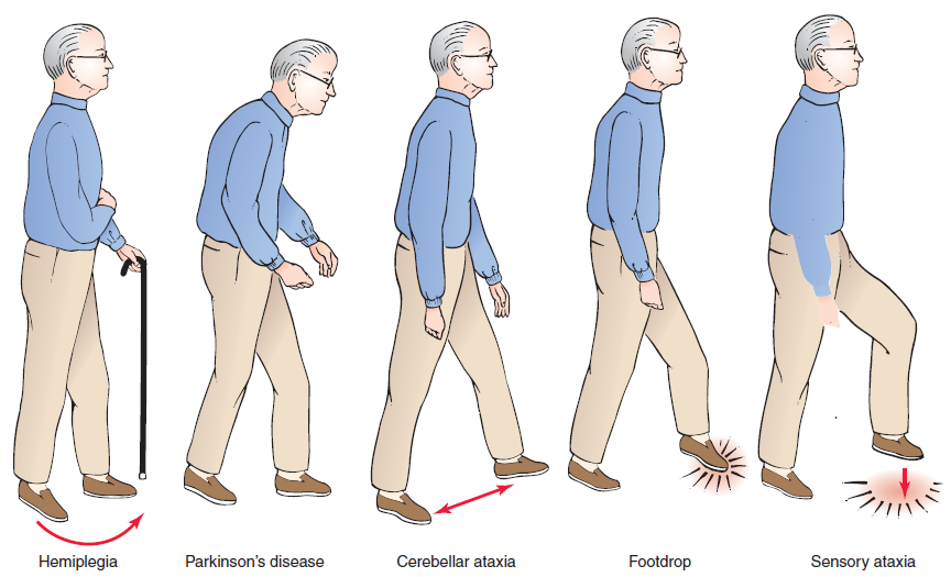
  • Gait disturbance (Figure 1)
  • Stooped posture, decreased arm swing
  • Difficulty initiating the first step; small shuffling steps that increase in speed (festinating gait). Steps become progressively faster and shorter while the trunk inclines farther forward.
  • Other early complaints and findings include micrographia (handwriting becomes smaller) and hypophonia (voice becomes softer).
  • Figure 1: Common types of gait abnormalities
  • Postural instability—tested by the pull test. Ask patient to stand in place with his or her back to the examiner. The examiner pulls the patient back by the shoulders; the proper response would be to take no or very few steps back without falling. Retropulsion is a positive test, as is falling straight back. This is not usually severe early. If falls and postural refl exes are greatly impaired early, then consider other disorders.
CAUSES
  • Unknown
  • Most cases are sporadic, with age being the most common risk factor, although there is probably a combination of environmental and genetic factors contributing to disease expression. There are rare familial forms with at least five different genes identified. The most well known is the parkin gene, which is a significant cause of early-onset autosomal recessive Parkinson’s disease and isolated juvenile-onset Parkinson’s disease (at or before age 20).
DIAGNOSIS
  • A presumptive clinical diagnosis can be made based on a comprehensive history and physical examination. The combination of asymmetrical signs, resting tremor, and good response to levodopa best differentiates idiopathic Parkinson’s disease from other causes of parkinsonism (see “Differential Diagnosis” for PD).
DIFFERENTIAL DIAGNOSIS
  • Multisystem atrophy—distinguishing features include autonomic dysfunction, (including urinary incontinence, orthostatic hypotension, and erectile dysfunction), parkinsonism, cerebellar signs, and normal cognition.
  • Diffuse Lewy body disease—parkinsonism with concomitant dementia. Patients often have early hallucinations and fluctuations in levels of alertness and mental status.
  • Corticobasal degeneration—often begins asymmetrically with apraxia, cortical sensory loss in one limb, and sometimes alien limb phenomenon.
  • Progressive supranuclear palsy—tends to have axial rigidity greater than appendicular (limb) rigidity. These patients have early and severe postural instability. A hallmark is supranuclear gaze palsy that usually involves vertical gaze before horizontal.
  • Essential tremor—bilateral postural and action tremor.
  • Secondary (acquired) parkinsonism
    • Postinfectious parkinsonism—von Economo’s encephalitis
    • Parkinson’s pugilistica—after repeated head trauma
    • Iatrogenic—any of the neuroleptics and antipsychotics. The high potency D2-blocker neuroleptics are most likely to cause parkinsonism.
    • Toxins (e.g., MPTP, manganese, carbon monoxide)
  • Cerebrovascular disease (basal ganglia infarcts)
IMAGING STUDIES
  • CT has almost no role in investigations. MRI of the head may sometimes distinguish between idiopathic Parkinson’s disease and other conditions that present with signs of parkinsonism.
REFERENCE
  • For management and treatment please read full information:
    • Ferri's Clinical Advisor
    • American Family Physicians Journal
    • The Washington Manual 
    • Medscape Reference
    • The New England Journal of Medicine

Friday, September 5, 2014

Gastric Cancer

PATIENT HISTORY

A 72-year-old Japanese immigrant was brought in by his family with complaints of difficulty in eating, vague abdominal pain, and weight loss. Endoscopy and biopsy confirmed gastric adenocarcinoma (Figure1). Liver metastases were found on abdominal CT. The family and the patient chose only comfort measures and the patient died 6 months later.

Figure 1. Endoscopy show a raise and irregular
mass in the antrum of the stomach deforming the pylorus
INTRODUCTION

Gastric cancer is a malignant neoplasm of the stomach, usually adenocarcinoma.


ETIOLOGY AND PATHOPHYSIOLOGY

  • Eighty-five percent of stomach cancers are adenocarcinomas with 15% lymphomas and GI stromal tumors. Adenocarcinoma is further divided into two types:
    • Diffuse type—Characterized by absent cell cohesion, these tumors affect younger individuals infiltrating and thickening the stomach wall; the prognosis is poor. Several susceptibility genes have been identified for this type of cancer.
    • Intestinal type—Characterized by adhesive cells forming tubular structures, these tumors frequently ulcerate.
  • Tumor grade can be well (4.1%), moderate (23.1%), or poorly differentiated (54.9%), or undifferentiated (2.9%) (SEER data from 1988–2001; unknown type accounted for 15%).
  • Most tumors are thought to arise from ingestion of nitrates that are converted by bacteria to carcinogens. Exogenous and endogenous factors (see “Risk Factors” below) contribute to this process.
    • Exogenous sources of nitrates—Sources include foods that are dried, smoked, and salted. Helicobacter pylori infection may contribute to carcinogenicity by creating gastritis, loss of acidity, and bacterial growth
    • Oncogenic pathways identified in most gastric cancers are the proliferation/stem cell, nuclear factor-κB, and Wnt/β-catenin; interactions between them appear to influence disease behavior and patient survival.
    • Gastric tumors are classified for staging using the T (tumor) N (nodal involvement) M (metastases) system. Two important prognostic factors are depth of invasion through the gastric wall (less than T2 [tumor invades muscularis propria]) and presence or absence of regional lymph node involvement (N0). Changes made to the classification system in the seventh edition of the American Joint Commission’s Cancer Staging Manual for gastric cancer demonstrate better survival discrimination.
    • Gastric cancer spreads in multiple ways:
      • Local extension through the gastric wall to the perigastric tissues, omenta, pancreas, colon, or liver
      • Lymphatic drainage through numerous pathways leads to multiple nodal group involvement (e.g., intraabdominal, supraclavicular) or seeding of peritoneal surfaces with metastatic nodules occurring on the ovary, periumbilical region, or peritoneal cul-de-sac.
      • Hematogenous spread is also common with liver metastases.
RISK FACTORS
  • Previous gastric surgery—As a result of alteration of the normal pH or with biopsy showing high-grade dysplasia.
  • Other endogenous risk factors—Atrophic gastritis (including postsurgical vagotomized patients) and pernicious anemia are conditions that favor the growth of nitrate-converting bacteria. In addition, intestinal-type cells that develop metaplasia and possibly atypia can replace the gastric mucosa in these patients. Genetic polymorphisms (e.g., interleukin-1B-511, interleukin-1RN, and tumor necrosis factor-α) also appear to play a role. Familial adenomatous polyposis and hereditary nonpolyposis colorectal cancer are also risk factors.
  • Individuals infected with certain H. pylori bacteria (cytotoxinassociated gene A) are at increased risk of gastric adenocarcinoma (especially noncardia) and gastric mucosa-associated lymphoid tissue (MALT) lymphoma.
  • Additional risk factors—Smoking, low socioeconomic class, lower educational level, exposure to certain pesticides (e.g., those who work in the citrus fruit industry in fields treated with 2,4-dichlorophenoxyacetic acid [2,4-D], chlordane, propargite, and triflurin), radiation exposure, and blood type A.
DIAGNOSIS
  • Clinical features
    • Asymptomatic, if superficial and/or early.
    • Upper abdominal pain that ranges from vague to severe.
    • Postprandial fullness.
    • Anorexia and mild nausea are common
    • Nausea and vomiting occur with pyloric tumors.
    • Late symptoms include weight loss and a palpable mass (regional extension).
    • Late complications include peritoneal and pleural effusions; obstruction of the gastric outlet; bleeding from esophageal varices or postsurgical site; and jaundice.
    • Physical signs are also late features and include:
      • Palpable enlarged stomach with succussion splash (splashing sound on shaking, indicative of the presence of fluid and air in a body cavity).
      • Primary mass (rare).
      • Enlarged liver.
      • Enlarged, firm to hard, lymph nodes (i.e., left supraclavicular [Virchow]), periumbilical region (Sister Mary Joseph node), and peritoneal cul-de-sac (Blumer shelf; palpable on vaginal or rectal examination).
IMAGING AND ENDOSCOPY
  • Diagnosis can be made on endoscopy (Figures 1 and 2) with biopsy of suspicious lesions. Confocal laser endomicroscopy may improve detection of early lesions.
  • Urgent referral for endoscopy (within 2 weeks) is recommended for patients with dyspepsia who also have GI bleeding, dysphagia, progressive unexplained weight loss, persistent vomiting, irondeficiency anemia, epigastric mass, family history of gastric cancer (onset <50 years), or whose dyspepsia is persistent and they are older than age 55 years.
  • Double-contrast radiography is an alternative to endoscopy and can detect large primary tumors but distinguishing benign from malignant disease is difficult.
  • Although endoscopy is not necessary when radiography demonstrates a benign-appearing ulcer with evidence of complete healing at 6 weeks, some authors recommend routine endoscopy, biopsy, and brush cytology when any gastric ulcer is identified.
  • Some gastric polyps (adenomas, hyperplastic) have malignant potential and should be removed.
  • Work-up for metastases includes:
    • Chest radiograph
    • CT scan or MRI of the abdomen and pelvis
    Figure 2: Endoscopy showing a deep ulcer with
    yellow-brown exudate in the center of the mass,
    consistent with cancer.
  • Endoscopic sonography is useful as a staging tool when the CT scan fails to find evidence of locally advanced or metastatic disease.
LABORATORY STUDIES
  • A hemoglobin or hematocrit can identify anemia, present in approximately 30% of patients.
  • Electrolyte panels and liver function tests can assist in assessing the patient’s clinical state and any liver involvement.
  • Carcinoembryonic antigen (CEA) is increased in about half of cases.
DIFFERENTIAL DIAGNOSIS
  • Peptic ulcer—Typical symptoms include epigastric pain (described as a gnawing or burning), occurring 1 to 3 hours after meals and relieved by food or antacids. Patients may also have nausea and vomiting, bloating, abdominal distention, and anorexia. Endoscopy confirms diagnosis ( See Peptic Ulcer Disease )
  • Nonulcer dyspepsia—Includes gastroesophageal reflux disease and functional dyspepsia. Classic symptoms of gastroesophageal reflux disease are heartburn (i.e., substernal pain that may be associated with acid regurgitation or a sour taste) aggravated by bending forward or lying down, especially after a large meal; individual symptoms, however, do not help to distinguish these patients from those with peptic ulcer disease. Endoscopy is considered if symptoms fail to respond to treatment (e.g., histamine-2 receptor agonist, proton pump inhibitor) or red flag signs/symptoms occur (e.g., bleeding, dysphagia, severe pain, weight loss).
  • Chronic gastritis—Includes autoimmune (body-predominant) and H. pylori-related (antral-predominant) types; mucosal inflammation (primarily lymphocytes) may progress to atrophy and metaplasia. Abdominal pain and dyspepsia are common symptoms and patients may have pernicious anemia.
  • Esophagitis—May be mechanical or infectious (primarily viral and fungal). Symptoms include heartburn (retrosternal wave-like pain that may radiate to the neck or jaw) and painful swallowing (odynophagia); regurgitation of sour or bitter tasting material may occur with obstruction. Barium swallow or esophagoscopy can be used to establish the diagnosis.
  • Esophageal cancer—Relatively uncommon malignancy of two cell types: squamous cell cancers (largely related to smoking, excessive alcohol consumption, and other agents causing mucosal trauma) and adenocarcinomas (usually arising in the distal esophagus related to reflux disease). Symptoms include progressive dysphagia and weight loss; the diagnosis is confirmed on esophagoscopy and biopsy.
REFERENCE
  • For Management and Treatment Please Read Full Information
    • Ferri's Clinical Advisor
    • American Family Physician Journal 
    • The Washington Manual 
    • Medscape
    • The New England Journal of Medicine
    • The Journal of American Medical Association

Peptic Ulcer Disease

PATIENT STORY

A 41-year-old man presents with a 4-month history of epigastric pain. The pain is dull, achy, and intermittent; there is no radiation of the pain and it has not changed in character since it began. Coffee intake seems to exacerbate the symptoms while eating or drinking milk helps. Infrequently, he is awakened at night from the pain. He reports no weight loss, vomiting, melena, or hematochezia. On examination, there is mild epigastric tenderness with no rebound or guarding. The reminder of the examination is unremarkable. A stool antigen test is positive for Helicobacter pylori, and the patient is treated for peptic ulcer disease with eradication therapy.

INTRODUCTION

Peptic ulcer disease (PUD) is a disease of the gastrointestinal (GI) tract characterized by a break in the mucosal lining of the stomach or duodenum secondary to pepsin and gastric acid secretion, this damage is greater than 5 mm in size and with a depth reaching the submucosal layer.

EPIDEMIOLOGY

  • PUD is a common disorder affecting approximately 4.5 million people annually in the United States. It encompasses both gastric and duodenal ulcers (Figures 1 and 2).
  • One-year point prevalence is 1.8%, and the lifetime prevalence is 10% in the United States
  • Prevalence is similar in both sexes, with increased incidence with age. Duodenal ulcers most commonly occur in patients between the ages of 30 and 55 years, whereas gastric ulcers are more common in patients between the ages of 55 and 70 years.
  • PUD incidence in H. pylori-infected individuals is approximately 1% per year (6- to 10-fold higher than uninfected subjects).
  • Physician office visit and hospitalization for PUD have decreased in the last few decades.
  • The current U.S. annual direct and indirect health care costs of PUD are estimated at approximately $10 billion. However, the incidence of peptic ulcers keeps declining, possibly as a result of the increasing use of proton pump inhibitors and eradication of H. pylori infection.
ETIOLOGY AND PATHOPHYSIOLOGY
  • Causes of PUD include:
    • NSAIDs, chronic H. pylori infection, and acid hypersecretory states such as Zollinger-Ellison syndrome
    • Uncommon causes include Cytomegalovirus (especially in transplantation recipients), systemic mastocytosis, Crohn disease, lymphoma, and medications (e.g., alendronate).
    • Up to 10% of ulcers are idiopathic.
  • Infection with H. pylori, a short, spiral-shaped, microaerophilic Gram-negative bacillus, is the leading cause of PUD. It is associated with up to 70% to 80% of duodenal ulcers.
  • H. pylori colonize the deep layers of the gel that coats the mucosa and disrupt its protective properties causing release of certain enzymes and toxins. These make the underlying tissues more vulnerable to damage by digestive juices and thus cause injury to the stomach (Figures 59-1 to 59-3) and duodenum cells.
  • NSAIDs are the second most common cause of PUD and account for many H. pylori-negative cases.
  • NSAIDs and aspirin inhibit mucosal cyclooxygenase activity reducing the level of mucosal prostaglandin causing defects in the protective mucous layer.
  • There is a 10% to 20% prevalence of gastric ulcers and a 2% to 5% prevalence of duodenal ulcers in long-term NSAID users. The annual risk of a life-threatening ulcer-related complication is 1% to 4% in long-term NSAID users, with older patients having the highest risk.
RISK FACTORS
  • Severe physiologic stress—Burns, central nervous system trauma, surgery, and severe medical illness increase the risk for secondary (stress) ulceration.
  • Smoking—Evidence that tobacco use is a risk factor for duodenal ulcers in not conclusive, with several studies producing contradictory findings. However, smoking in the setting of H. pylori infection may increase the risk of relapse of PUD.
  • Alcohol use—Ethanol is known to cause gastric mucosal irritation and nonspecific gastritis. Evidence that consumption of alcohol is a risk factor for duodenal ulcer is inconclusive.
  • Medications—Corticosteroids alone do not increase the risk for PUD; however, they can potentiate ulcer risk in patients who use NSAIDs concurrently.
DIAGNOSIS

CLINICAL FEATURES
  • Epigastric pain (dyspepsia), the hallmark of PUD, is present in 80% to 90% of patients; however, this symptom is not sensitive or specific enough to serve as a reliable diagnostic criterion for PUD. Pain is typically described as gnawing or burning, occurring 1 to
    3 hours after meals and relieved by food or antacids. It can occur at night, and sometimes radiates to the back. Less than 25% of patients with dyspepsia have ulcer disease at endoscopy.
  • Other dyspeptic symptoms including belching, bloating, and distention are common but also not specific features of PUD as they are commonly encountered in many other conditions.
  • Additional symptoms include fatty food intolerance, heartburn, and chest discomfort.
  • Nausea and anorexia may occur with gastric ulcers.
  • Significant vomiting and weight loss are unusual with uncomplicated ulcer disease and suggest gastric outlet obstruction or gastric malignancy.
  • Twenty percent of patients with ulcer complications such as bleeding and nearly 61% of patients with NSAID-related ulcer complications have no antecedent symptoms.
  • Rare and nonspecific physical findings include:
    • Epigastric tenderness
    • Heme-positive stool
    • Hematemesis or melena in cases of GI bleeding
TYPICAL DISTRIBUTION
  • Duodenal ulcers occur most often in the first portion of the duodenum (>95%), with approximately 90% of ulcers located within 3 cm of the pylorus.
  • Benign gastric ulcers are located most commonly in the antrum (60%) and at the junction of the antrum and body on the lesser curvature (25%) (Figure 3).
LABORATORY STUDIES
  • In most patients with uncomplicated PUD, routine laboratory tests are not helpful
  • Noninvasive tests include serum H. pylori antibody detection, fecal antigen tests, and urea breath tests; the latter two, if positive, indicate active disease.
  • Serum enzyme-linked immunosorbent assay (ELISA) is the least accurate test and is useful only for diagnosing the initial infection.
  • The stool antigen test is less convenient but is highly accurate and also can be used to confirm H. pylori eradication, as can the urea breath test.
  • Obtaining a serum gastrin may be useful in patients with recurrent, refractory, or complicated PUD and in patients with a family history of PUD to screen for Zollinger-Ellison syndrome.
  • Barium upper gastrointestinal (UGI) series is an acceptable alternative to endoscopy but is not as sensitive for the diagnosis of small ulcers (<0.5 cm) and does not allow for biopsy with gastric ulcer.
  • Patient’s testing positive for PUD should undergo noninvasive testing for H. pylori.
  • UGI series has limited accuracy in distinguishing benign from malignant gastric ulcers; therefore, all patients diagnosed this way should be reevaluated with endoscopy after 8 to 12 weeks of therapy.
DIFFERENTIAL DIAGNOSIS

Disease processes that may present with “ulcer-like” symptoms include:
  • Nonulcer or functional dyspepsia (FD)—The most common diagnosis among patients seen for upper abdominal discomfort; it is a diagnosis of exclusion. Dyspepsia has been reported to occur in up to 30% of the U.S. population.
  • Gastroesophageal reflux—Classic symptoms are heartburn (i.e., substernal pain that may be associated with acid regurgitation or a sour taste) aggravated by bending forward or lying down, especially after a large meal. Endoscopy is considered if symptoms fail to respond to treatment (e.g., histamine-2-receptor agonist, proton pump inhibitor [PPI]) or red flag signs and symptoms occur.
  • Gastric cancer—Most patients do not become symptomatic until late in the disease; symptoms include upper abdominal pain, postprandial fullness, anorexia and mild nausea, vomiting (especially with pyloric tumors), weight loss and a palpable mass. Endoscopic biopsy is used
    to make this diagnosis.
  • Biliary colic is characterized by discrete, intermittent episodes of pain that should not be confused with other causes of dyspepsia.
  • Gastroduodenal Crohn disease—Symptoms include epigastric pain, nausea, and vomiting. On endoscopy, patients often have H. pylorinegative gastritis and may develop gastric outlet obstruction. Extraintestinal manifestations include erythema nodosum, peripheral
    arthritis, conjunctivitis, uveitis, and episcleritis. Endoscopy shows an inflammatory process with skip lesions, fistulas, aphthous ulcerations, and rectal sparing. Small bowel involvement is seen on imaging with longitudinal and transverse ulceration (cobblestoning) in addition to segmental colitis and frequent stricture.
MANAGEMENT
  • The approach to patients with dyspepsia includes performing endoscopy for patients with red flag symptoms or who are older than age 55 years. For patients who have an ulcer identified on
    endoscopy, eradication of H. pylori is attempted (as below) and a PPI is continued for 4 to 8 weeks. For those without an ulcer on endoscopy, treatment with a PPI or H2 blocker is provided.
  • For patients without red flag findings, testing and treating for H. pylori; counseling to avoid smoking, alcohol, and NSAIDs; and appropriate use of antisecretory therapy for 4 weeks will be successful in the majority of patients.
  • The goals of treatment of active H. pylori-associated ulcers are to relieve dyspeptic symptoms, to promote ulcer healing, and to eradicate H. pylori infection. Eradication of H. pylori is better than ulcer-healing drug therapy for duodenal ulcer healing8 and greatly reduces the incidence of ulcer recurrence from 67% to 6% in patients with duodenal ulcers and from 59% to 4% in patients with gastric ulcers.
  • The worldwide empiric use of traditional triple therapy with PPI, clarithromycin, and amoxicillin no longer provides an acceptable cure rate (cure rate below 80%) because of the increasing prevalence of clarithromycin resistance.
  • Four drug combinations currently provide the best results and consist of two general combinations: (a) a PPI, amoxicillin, clarithromycin, metronidazole/tinidazole given either sequentially or concomitantly, or (b) a PPI, a bismuth, tetracycline HCL, and metronidazole/tinidazole.
  • A PPI, levofloxacin, and amoxicillin for 10 days appears to be more effective and better tolerated than a PPI, bismuth, tetracycline, and metronidazole in patients with persistent H. pylori infection but requires validation in North America.
  • The European Helicobacter Study Group consensus guideline states that triple therapy (PPI-clarithromycin-amoxicillin/metronidazole) or Bismuth quadruple therapy remain first-line treatment in areas with low clarithromycin resistance. For areas with high clarithromycin
    resistance (>20%), Bismuth or non-Bismuth quadruple therapy are preferred.
  • Treat NSAID-induced ulcers with cessation of NSAIDs, if possible, and an appropriate course of standard ulcer therapy with a H2-receptor antagonist or a PPI. If NSAIDs are continued, prescribe a PPI.
  • H. pylori-negative ulcers that are not caused by NSAIDs can be treated with appropriate antisecretory therapy, either H2-receptor antagonist or PPI.
  • For patients with bleeding peptic ulcers, high-dose PPIs do not reduce rates of rebleeding, surgical intervention, or mortality after endoscopic treatment compared with non–high-dose PPIs.
FOLLOW-UP
  • Endoscopy is required to document healing of gastric ulcers and to rule out gastric cancer; this is performed 6 to 8 weeks after the initial diagnosis.
  • Confirmation of H. pylori eradication in patients with uncomplicated ulcers is not necessary.
  • Confirmation of healing with endoscopy is required in all patients with ulcer complicated by bleeding, perforation, or obstruction.
  • For patients without initial endoscopy who have persistent symptoms following initial treatment, the PPI or H2 blocker can be continued for another 4 to 8 weeks.7 If there is inadequate response to therapy, endoscopy and evaluation for hypersecretory states should
    be considered.
Figure 1: Endoscopic pictures of a gastric ulcer.

Figure 2: Endoscopic view of a pyloric ulcer and
an erosion of the mucosa.

Figure 3: Stomach ulcer in a patient with a hiatal hernia.


REFERENCES


For Management and Treatment Please Check These:

  • Ferri's Clinical Advisor
  • American Family Physician Journal 
  • The Washington Manual 
  • Medscape
  • The New England Journal of Medicine
  • The Journal of American Medical Association