The steps in one complete circuit through the cardiovascular system are shown in figure. The cycled numbers in the figure correspond with the steps described here:
 |
| A schematic diagram showing the circuitry of the cardiovascular system |
- Oxygenated blood fills the left ventricle. Blood that has been oxygenated in the lungs returns to the left atrium via the pulmonary vein. This blood then flows from the left atrium to the left ventricle through the mitral valve (the AV valve of the left heart).
- Blood is ejected from the left ventricle into the aorta. Blood leaves the left ventricle through the aortic valve (the semilunar valve of the left side of the heart), which is located between the left ventricle and the aorta. When the left ventricle contracts, the pressure in the ventricle increases, causing the aortic valve to open and blood to be ejected forcefully into the aorta. (As noted previously, the amount of blood ejected from the left ventricle per unit time is called the cardiac output.) Blood then flows through the arterial system, driven by the pressure created by contraction of the left ventricle.
- Cardiac output is distributed among various organs. The total cardiac output of the left heart is distributed among the organ systems via sets of parallel arteries. Thus, simultaneously, 15% of the cardiac output is delivered to the brain via the cerebral arteries, 5% is delivered to the heart via the coronary arteries, 25% is delivered to the kidneys via the renal arteries, and so forth. Given this parallel arrangement of the organ systems, it follows that the total systemic blood flow must equal the cardiac output.
- Blood flow from the organs is collected in the veins. The blood leaving the organs is venous blood and contains waste products from metabolism, such as carbon dioxide (CO2). This mixed venous blood is collected in veins of increasing size and finally in the largest vein, the vena cava. The vena cava carries blood to the right heart.
- Venous return to the right atrium. Because the pressure in the vena cava is higher than in the right atrium, the right atrium fills with blood, the venous return. In the steady state, venous return to the right atrium equals cardiac output from the left ventricle.
- Mixed venous blood fills the right ventricle. Mixed venous blood flows from the right atrium to the right ventricle through the AV valve in the right heart, the tricuspid valve.
- Blood is ejected from the right ventricle into the pulmonary artery. When the right ventricle contracts, blood is ejected through the pulmonic valve (the semilunar valve of the right side of the heart) into the pulmonary artery, which carries blood to the lungs. Note that the cardiac output ejected from the right ventricle is identical to the cardiac output that was ejected from the left ventricle. In the capillary beds of the lungs, oxygen (O2) is added to the blood from alveolar gas, and CO2 is removed from the blood and added to the alveolar gas. Thus, the blood leaving the lungs has more O2 and less CO2 than the blood that entered the lungs.
- Blood flow from the lungs is returned to the heart via the pulmonary vein. Oxygenated blood is returned to the left atrium via the pulmonary vein to begin a new cycle.
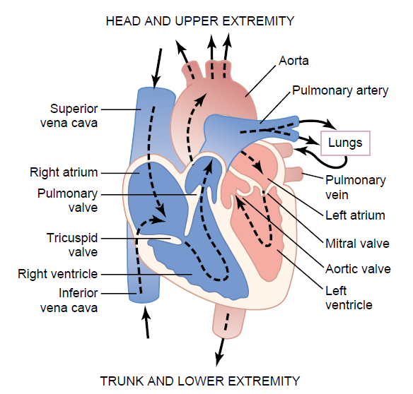 |
| Structure of the heart, and course of blood flow through the heart chambers and heart valves. |


0 comments:
POST A COMMENT